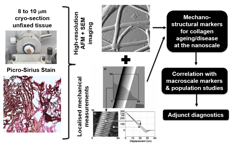
We are developing a “quantitative nano-histology (QNH)” approach that can be applied to clinical practice with regards to diagnosis, staging and prognostication of Head and Neck Squamous Cell Carcinoma (as one example).
QNH refers to the combination sub-micron structural imaging and mechanical analysis (by Atomic Force Microscopy, or AFM) performed directly on histological sections (unfixed – fresh with or without immuno-staining).
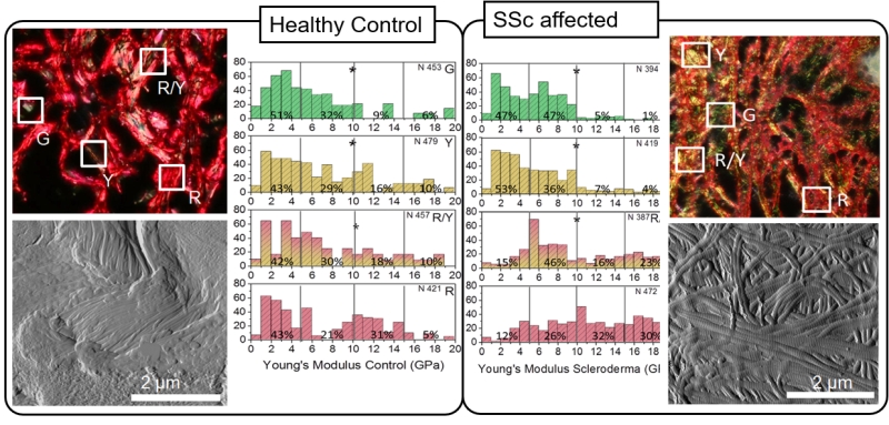
Using an in-house developed approach, we have been able to define dermal structural and mechanical markers as a function of aging and skin fibrosis.
While we understand that AFM may constitute a robust and objective way to analyze histological samples for surface characterization as a way to complement standard histological procedures for skin, in particular, further research in the assessment of cancer tissue nanotopography by ultrastructure imaging of histological blocks using AFM is required.
This approach is currently being expanded collaboratively to collagen fibrosis and connective disorders such as Elhers Danlos.
Related published papers
Ahmed, T., Nash, A., Clark, K. E., Ghibaudo, M., de Leeuw, N. H., Potter, A. … & Bozec, L. (2017). Combining nano-physical and computational investigations to understand the nature of “aging” in dermal collagen. International Journal of Nanomedicine, 12, 3303–3314.
Strange, A. P., Aguayo, S., Ahmed, T., Mordan, N., Stratton, R., Porter, S. R. & Bozec, L. (2017). Quantitative nanohistological investigation of scleroderma: An atomic force microscopy-based approach to disease characterization. International Journal of Nanomedicine, 12, 411-420.